Dentist in Aurora: The Importance of Regular X-Rays
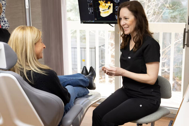
Dentistry relies on light and line of sight, and the mouth does not always cooperate. Teeth sit shoulder to shoulder. Gums and cheeks cast shadows. Early decay, hairline fractures, abscesses, infections buried in bone, even extra teeth can hide in places a mirror cannot reach. That is why regular dental X-rays are not an optional add-on, they are part of accurate diagnosis and safe, conservative care. If you see a dentist in Aurora for routine checkups, you have likely had X-rays taken at some point. Understanding what they show, how often you need them, and why they are safe helps you make better decisions for yourself and your family.
I have sat with parents who felt uneasy about radiation for their six-year-old, and with athletes who waited out a toothache until an X-ray revealed a deep abscess inches from their sinus. I have also seen a spotless mouth on visual exam, then found between-tooth cavities on a bitewing series the same day. Experience teaches the same lesson over and over: the most expensive dentistry is the problem that was missed early. Well-timed images prevent that.
What X-rays reveal that a mirror cannot
Most dental disease begins in tight spaces. Decay starts in the grooves of molars and creeps into the contact points where brushes do not reach. Periodontal disease thins the bone that anchors teeth, long before gums look dramatically different. Cysts, impacted canines, and root problems develop within the jawbone, far from any visible surface. Without imaging, a dentist is guessing about the three dimensional reality behind a two dimensional view.
Here are a few patterns that come up in everyday practice:
A patient with occasional cold sensitivity on a back tooth might have a small interproximal cavity between molars. The enamel still looks intact to the eye, but a bitewing X-ray shows the telltale triangle of demineralization. A quick filling prevents a root canal later.
A teenager with crowding and a tiny gap near a canine can look routine. A panoramic X-ray sometimes shows that the canine is actually pointed horizontally and on a collision course with the lateral incisor root. Intervening in middle school can save both teeth.
A person with persistent gum bleeding, despite good home care, may be experiencing early bone loss. Bitewings are sensitive to changes in bone height. Seeing that pattern defines the difference between a simple cleaning and a targeted periodontal plan.
Cracks in heavily restored molars can be elusive. A periapical image, combined with a careful bite test, often shows a dark line at the root tip or a widened ligament space that betrays a split. Catching the crack early may allow for a crown, instead of waiting for a vertical fracture that forces extraction.
These are the quiet problems regular X-rays help catch before they become noisy emergencies.
Frequency: how often do you need dental X-rays?
There is no one size fits all schedule, and that is a good thing. A dentist in Aurora will tailor imaging to your risk. Guidance from professional organizations supports this individualized approach with ranges rather than rules.
For children with mixed dentition and average cavity risk, bitewings are often taken every 6 to 12 months. The enamel on baby teeth is thinner and lesions progress faster, so waiting two years can mean missing a problem that went from small to big.
Teenagers with braces tend to carry higher risk because brackets complicate hygiene. Bitewings roughly every 6 to 12 months, plus a panoramic X-ray to monitor developing roots and impacted teeth, is typical. The panoramic is not annual by default, usually every few years or when there is a change in alignment or symptoms.
Healthy adults with little to no history of decay and excellent home care may need bitewings every 18 to 24 months. If you have a run of cavity free checkups, fluoride exposure through your water or products, and minimal plaque, stretching intervals can be reasonable.
Adults with risk factors, such as dry mouth from medications, diabetes, frequent snacking, tobacco use, or a history of frequent cavities, benefit from more frequent bitewings, about every 6 to 12 months. The same applies if you have many older fillings or crowns that can develop recurrent decay at the margins.
New patients, regardless of age, usually need a baseline set of images to establish a complete record. This often includes bitewings plus selected periapicals, or a panoramic X-ray. If you can transfer recent images from a previous Dental clinic in Aurora or another city and they are diagnostically useful, your new dentist may defer retaking them.
Think of frequency as an outcome of your risk profile and recent findings, not a calendar checkbox. When the mouth is quiet and your risk is low, your dentist can reasonably space out imaging. When something changes, more information protects you.
Types of dental X-rays and what each one shows
-
Bitewings: Focused images that capture the crowns of upper and lower teeth together, usually in the back of the mouth. Best for spotting cavities between teeth and assessing bone levels around molars and premolars. Typically taken in sets of two or four.
-
Periapicals: Close up images that include the full tooth from crown to root tip, plus the surrounding bone. Ideal for diagnosing abscesses, root issues, cracks, and problems under or around a single tooth. Often used when a specific tooth hurts.
-
Panoramic: A broad, single image of the upper and lower jaws, sinuses, TMJ regions, and developing teeth. Useful for evaluating impacted teeth, jaw joints, cysts, tumors, and overall anatomy. Common in orthodontic planning and for wisdom teeth.
-
Cone beam CT (CBCT): Three dimensional imaging used for complex cases such as implant placement, root canal retreatment, airway evaluation, or impacted canines. Provides depth and spatial relationships that 2D images cannot show. Not needed for routine checkups.
These categories cover the majority of clinical needs. A dentist in Aurora will choose the smallest field and lowest dose that still answers the clinical question, which brings us to safety.
Safety and dose: understanding the numbers
Radiation conversations work best with actual measurements, not vague reassurances. We measure dose in microsieverts, abbreviated µSv. Everyone receives background radiation every day from the sun, soil, and food. In most of North America, background averages around 3,000 µSv per year, which is about 8 to 10 µSv per day.
Modern digital dental X-rays are low dose, particularly with rectangular collimation and high speed sensors. Ranges vary by equipment and technique, but reasonable ballpark figures are:
A set of four digital bitewings: roughly 5 to 20 µSv. At the lower end with optimized settings, the dose is comparable to roughly one to two days of natural background radiation.
A single periapical: about 1 to 5 µSv.
A panoramic image: roughly 9 to 26 µSv, often in the range of two to three days of background radiation.
A small field CBCT scan: about 20 to 100 µSv, sometimes higher with larger fields. Even here, we are often talking about the equivalent of several days to a couple of weeks of background dose, not months.
For comparison, a medical chest X-ray is commonly around 100 µSv or more, depending on technique.
Dentistry follows the ALARA principle, as low as reasonably achievable, and more recently ALADAIP, as low as diagnostically acceptable, indication oriented, and patient specific. In practice this means using the narrowest beam, the fastest sensor, protective thyroid collars when appropriate, and exposure only when the diagnostic benefit justifies it. Current professional guidance notes that lead aprons are not essential with modern equipment, because the beam is tightly collimated to the head, but many offices still use aprons to reassure patients. There is nothing wrong with that added comfort, as dentist Aurora long as it does not interfere with the image.
If you are pregnant, it is common to defer non-urgent imaging, especially during the first trimester. That said, dental X-rays with shielding are considered safe when clinically necessary. Treating a painful infection promptly is safer for both parent and baby than letting it linger. Share your status with your dentist, and expect a discussion that weighs urgency, alternatives, and timing.
Special situations and how imaging guides care
Family dentistry in Aurora sees a range of needs in a typical week, from toddlers cutting their first molars to grandparents maintaining implant-supported dentures. Imaging protocols reflect these stages.
Young children benefit from early bitewings once their back teeth touch. Decay between baby molars can spread quickly and painlessly until it reaches the nerve. Two small bitewings often prevent a far more traumatic visit later.
Tweens and teens in orthodontic treatment need periodic panoramic images to watch root development and wisdom teeth. If canine teeth are off course, a targeted CBCT can map their position relative to neighboring roots, guiding a safe traction plan.
Athletes and grinders who clench at night are at risk for cracked teeth. Periapicals around suspect molars reveal changes at the tip of the roots that go hand-in-hand with a crack. Pairing that image with a bite stick test or transillumination helps decide if a crown, a root canal, or extraction is the right move.
Patients with periodontal disease rely on serial bitewings to measure bone height changes over time. Subtle improvements after scaling and root planing, or ongoing bone loss that needs surgical attention, do not always match what gums look like at a glance. The images provide the hard numbers.
People with dry mouth due to medications, head and neck radiation, autoimmune conditions, or simply aging, develop cavities at the gumline and between teeth at a faster clip. More frequent bitewings help intercept lesions when they are still small enough for conservative treatment.
Implant planning without 3D imaging is guesswork. A small field CBCT maps bone width, density, sinus position, and nerve location. That data informs whether grafting is needed, which implant size fits, and how to place it safely. For a single implant in an otherwise healthy patient, a limited field scan keeps dose modest while still delivering critical information.
Root canal specialists sometimes request a CBCT when a conventional retreatment fails or the anatomy is unusual. Extra canals can hide in upper molars and lower incisors. Three dimensional imaging prevents missed structures that could cause persistent infection.
What happens during a typical X-ray visit
Most people are surprised by how quick and uneventful modern imaging is. A hygienist or assistant places a small digital sensor or phosphor plate in your mouth, positions a lightweight aiming ring outside your cheek, and asks you to bite gently. The exposure takes a fraction of a second. If you have a sensitive gag reflex, request that upper molar images be taken first or ask for topical numbing gel on the palate. Breathing slowly through your nose and lifting one foot slightly off the chair during placement can distract the reflex just enough to get the image.
For a panoramic image, you stand or sit still while the machine rotates around your head. A bite block keeps your jaw in position. The whole sweep takes less than a minute. With CBCT, you hold still a bit longer, but the experience is similar, without the claustrophobia sometimes associated with medical CT scanners.
Afterward, your dentist reviews the images chairside, often zooming and adjusting contrast to point out details. Expect to see caries as darker areas within the lighter enamel, a thin white line around root tips when healing is underway, or a widened dark halo at a root tip when an infection is active. Good communication here matters. A dentist should correlate what you feel, what they see clinically, and what the image shows before proposing treatment.
A quick checklist: when X-rays are likely recommended
- You are a new patient without recent transferable images.
- You have tooth pain, sensitivity to biting, or swelling near a tooth.
- It has been 12 to 24 months since your last bitewings, depending on risk.
- Your child’s back teeth are touching and you want to prevent hidden cavities.
- You are planning implants, orthodontics, or wisdom tooth removal.
These are common triggers, not absolutes. A trusted dentist in Aurora will discuss the why behind each image.
Trade-offs, costs, and when to defer
Prudent imaging protects you from both overtreatment and undertreatment. That balance includes cost and radiation, and it respects your preferences.
If you had bitewings taken at a Dental clinic in Aurora three months ago and you can obtain copies, there is rarely a reason to retake them unless there is a new symptom. Digital files transfer easily. Most offices are happy to share when you sign a release.
Insurance plans often allow bitewings once per year or once every 18 months, with a panoramic every three to five years. Those rules do not always match clinical reality. If you fall between benefits but have clear indications, you can still choose imaging and pay out of pocket. Conversely, if benefits would cover an image that is not clinically needed, a conscientious dentist should still say, not today.
There are times to wait. If you are pregnant and the problem is minor and not painful, deferring until the second trimester or postpartum is reasonable. If you are a very low risk adult with stable exams for years, spacing bitewings to 24 months can make sense. Good dentistry is not about taking every possible picture, it is about taking the right picture at the right time.
Local perspective: finding the right fit in Aurora
Aurora is a city of families on the move, students juggling part-time jobs, healthcare workers keeping odd hours, and retirees with more time to travel than to sit in a waiting room. A Dental clinic in Aurora that respects that pace will build imaging into efficient visits. Look for practices that explain their rationale in plain language, use digital sensors, protect your thyroid when indicated, and adjust intervals to your risk rather than a fixed template.
Family dentistry in Aurora often serves multiple generations under one roof. That continuity lets your dentist compare your child’s bitewings year over year, or line up your periodontal bone levels across a decade. Subtle trends become obvious when the same eyes track them. If you change offices, bring your images with you. That continuity belongs to you.
Small stories from the chair
A middle school soccer player came in after a routine cleaning flagged nothing alarming. Mom almost declined bitewings because her son never had a cavity. The images showed early decay between the lower molars on both sides, still confined to enamel. Two tiny fillings later, he was back on the field. If they had waited a year, those spots could have crossed into dentin, requiring larger restorations and possibly a future crown.
A 42 year old nurse felt a dull ache near an upper molar that flared during flights. No visible decay, no broken cusp. A periapical X-ray revealed a periapical radiolucency at the palatal root, consistent with a dying nerve. An endodontic referral handled the root canal within the week. The pressure pain during plane landings stopped on the next trip.
An 80 year old gentleman with well maintained crowns continued to lose attachment around his lower incisors despite careful cleanings. Serial bitewings over two years documented progressive bone loss. That objective evidence tipped the decision toward a small periodontal surgery that stabilized the area. He kept his front teeth and his confident smile for his granddaughter’s wedding.
These are the sorts of clinical calls where X-rays inform, not replace, judgment.
Myths that keep people from timely imaging
Radiation fear sits at the top. Numbers help, especially put alongside everyday exposures. A set of modern bitewings delivers a dose in the range of one to two days of natural background radiation. Flying cross country exposes you to more cosmic radiation than a single panoramic image. Dental teams operate equipment designed to limit scatter and direct the beam precisely. You can ask to see the settings and shielding anytime.
Another misconception aspenwooddental.com Dentist in Aurora is that X-rays hurt. The sensor or plate can feel bulky, especially near upper molars where the palate is sensitive, but the exposure itself is painless and fast. Techniques like using smaller sensors where appropriate, warming the plate in a gloved hand, and applying a dab of topical anesthetic make a big difference.
Some worry that X-rays always lead to treatment. In reality, they often do the opposite. I have used images to show that a dark spot on a visual exam is simply a stained pit, not decay, and to justify monitoring instead of drilling. I have also used them to prove that a five year old filling is still well sealed. Good imaging underpins conservative dentistry.
How regular X-rays support long term savings
Dental disease tends to follow a predictable cost curve. A small interproximal cavity addressed with a conservative filling is relatively inexpensive and preserves most of the tooth. If it progresses into the nerve, you are looking at a root canal, a core build up, and a crown. If it cracks below the gumline, extraction and an implant or bridge sit on the table. Each step up the ladder costs two to four times more than the one before it, not counting time off work and discomfort.
Regular bitewings give you the chance to intervene at the earliest possible stage. Periodontal images do the same for bone loss. Over five to ten years, that translates into fewer big procedures, fewer emergencies, and a mouth that ages more gracefully. Patients sometimes tell me they want to avoid any X-rays to save money. I understand the instinct. My experience says the opposite happens.
Working with your dentist to set a smart X-ray plan
If you live or work near Aurora, ask your dentist to walk you through a personalized interval. The conversation should cover your cavity history, diet, fluoride exposure, saliva flow, home care routine, orthodontic plans, and any systemic factors like diabetes. Expect to see your most recent images onscreen with comparisons to prior years. If your last few checkups were clean and your bitewings are unchanged, your Dentist in Aurora may push the next set a little further out. If your child’s first molars just erupted and are touching, the schedule may tighten briefly.
Come with questions. Are rectangular collimators in use? Do you have thyroid collars available? Can we use the smallest field CBCT if a 3D scan is necessary? Can you send my images to my specialist or to me directly? A transparent office will answer yes to most of these and explain when exceptions apply.
You are not buying pictures. You are buying clarity. The right X-rays, taken at the right times, make your care safer, more precise, and usually more affordable over the long haul.
The bottom line for Aurora patients
If you have been putting off a checkup because you are worried about X-rays, talk it through with a local practice that respects both science and your comfort. A well run Dental clinic in Aurora uses digital technology, shields judiciously, and never takes an image without a clear reason. Family dentistry in Aurora thrives on relationships built over years, and regular, well calibrated X-rays are a quiet but central part of that trust.
When your dentist recommends imaging, ask what they hope to find, what alternatives exist, and how the result will change your care. When the answer is clear, saying yes is one of the simplest ways to protect your teeth for decades to come.
Aspenwood Dental Associates and Colorado Dental Implant Center
Address: 2900 S Peoria St Ste C, Aurora, CO 80014, United States
Phone number: +13037314037
FAQ About Dentist Aurora
How can I fix my teeth if I don't have money?
If you have no money, the most effective way to fix your teeth is to visit a Federally Qualified Health Center (FQHC) or a dental school clinic. FQHCs offer care on a sliding scale based on your income, and dental schools provide heavily discounted treatments performed by students under licensed supervision.
How do you know if the dentist you found is a good dentist or not?
A great dentist prioritizes your long-term oral health, communicates clearly about treatment options and costs, and makes you feel comfortable. You can easily evaluate if a dentist is a good fit by assessing their communication style, clinical environment, and patient feedback.
How do poor people get their teeth fixed?
People with limited finances often get their teeth fixed by utilizing government-funded clinics, visiting university dental schools for discounted care, or relying on regional charitable events. These avenues provide essential treatments like cleanings, fillings, and extractions to those who cannot afford traditional dental costs.